Showing posts with label spine surgery. Show all posts

Wednesday, 10 February 2016

Most Advanced Slip Disc Treatment in India | Best Spine Hospital India


The spine is made up of many bones called vertebrae. These are roughly circular and between each vertebra is a 'disc'. The discs are made of strong 'rubber-like' tissue which allows the spine to be fairly flexible. A disc has a stronger fibrous outer part, and a softer jelly-like middle part called the nucleus pulposus. 

The spinal cord, which contains the nerves that come from the brain, is protected by the spine. Nerves from the spinal cord come out from between the vertebrae to take and receive messages to various parts of the body. 

Strong ligaments attach to the vertebrae. These give extra support and strength to the spine. Various muscles also surround, and are attached to, various parts of the spine. (The muscles and ligaments are not shown in the diagram below for clarity.) 

At what age can a slipped disc occur? 
A slipped disc in the lower back is most often seen between the ages of 30 and 50. In the cervical vertebrae around the neck, slipped discs are most often seen between the ages of 40 and 60. 

Who Gets A Slipped Disc? 
  

What Are The Symptoms Of A Slipped Disc? 

Back pain

The pain is often severe, and usually comes on suddenly. The pain is usually eased by lying down flat, and is often made worse if you move your back, cough, or sneeze. 

Nerve root pain (usually 'sciatica')

Nerve root pain is pain that occurs because a nerve coming from the spinal cord is pressed on ('trapped') by a prolapsed disc, or is irritated by the inflammation caused by the prolapsed disc. Although the problem is in the back, you feel pain along the course of the nerve in addition to back pain. Therefore, you may feel pain down a leg to the calf or foot. Nerve root pain can range from mild to severe, but it is often worse than the back pain. With a prolapsed disc, the sciatic nerve is the most commonly affected nerve. (The term 'sciatica' means nerve root pain of the sciatic nerve.) The sciatic nerve is a large nerve that is made up from several smaller nerves that come out from the spinal cord in the lower back. It travels deep inside the buttock and down the back of the leg. There is a sciatic nerve for each leg. 

Lumbago, or what is more commonly known to people as lower back pain , is one of the most widespread and prevalent spinal problems. The reason for such persistent back pain is any kind of pathological problem in the lumbar region of the spine, which is made up of the last five vertebrae.

Lumbar spine problems are most often caused by herniated inter-vertebral discs, anomalous developments on the vertebral bodies (osteophytes), which apply pressure on the spinal nerves that leads to narrowing of the spinal column around the spinal cord. The most important procedures followed in a lumbar Spine Surgery are decompression and fusion.

Decompression Lumbar Decompression spine surgery in India is performed to assuage pain caused by pinched nerves. The surgery is generally recommended in cases of spinal stenosis caused by thickened joints, loosened ligaments, bony growths, or disc herniation.

The various procedures used for Decompressing Lumbar spine are:•  Discectomy•  Laminotomy or Laminectomy•  Port Hole Decompression•  Foraminotomy or Foraminectomy•  Osteophyte removal•  Corpectomy

Fusion : Lumbar fusion surgeryin India has two individual approaches. The most common process used is the posterior approach, where the surgery is done from the back. The three main Posterior fusion techniques are:

•  Postero lateral gutter fusion surgery•  Posterior lumbar interbody fusion (PLIF) surgery•  Transforaminal lumbar interbody fusion (TLIF) surgeryThe anterior approach involves placing the bone directly into the section between the vertebrae where the shock-absorption disc had been situated.

Candidates

Lumbarspine surgery in India is indicated for people who have pain that extends (radiates) from the back to the buttocks or back of thigh, pain that interferes with daily activities, weakness of legs or feet, numbness of legs, feet, or toes, loss of bowel of bladder control, had physiotherapeutic & medication input, but that hasn't helped.

Expected ResultsMost patients can expect a dramatic and lasting improvement in their back and leg pain after the lumbar spinal surgery . Though the success rates are excellent in Lumbar surgery , it can be lower in patients who smoke, are overweight, have diabetes or other significant medical illnesses, have osteoporosis, or who have had radiation treatments that included the lower back. Good nutrition and slowly increasing activity (as recommended by your physician) in the recovery period can help achieve success.

RecoveryYou are likely to experience pain and discomfort after the surgery which will be controlled with pain killers prescribed by your surgeon. You would be advised to walk the same day or the next after the surgery to prevent any clot formation. You would also be advised to do breathing exercises after the lumbar surgery for better circulation of blood and decrease the risk of any lung problem. You would need to stay at the hospital for about 5-6 days after the surgery and can go back home with specific instructions to follow. Many people return to work in 3-4 weeks after the surgery. But you may or may not be allowed to do heavy work depending upon your specific condition.

Thursday, 10 September 2015

Minimally Invasive Surgery (MIS) for Spine Treatment - World's Class Spine Hospitals in India

The term "Minimally Invasive" in most cases, when describing a surgical procedure, refers to a technique using small incisions to gain access to the particular area of the body needing treatment. By avoiding a larger incision, there can be less force from the retractors on the surrounding tissues, less tissue dissection, and usually better preservation of the surrounding blood supply to the tissues in the operated area. 

Ideally, these factors lead to quicker healing and a recovery with less pain, although these differences have not been proven conclusively. For an "open" procedure (non-MIS), the surgical wound is larger and more dissection is required. Retraction on the muscles, when kept in place during a long procedure, has been shown to cause damage to these muscles, with some of these effects possibly permanent.

The comparison between minimally invasive and open surgery can be made when describing a lumbar fusion. This procedure typically has the patient lying face down during the surgery, with the work done on the back side (posterior). With an open case, the incision is usually in the midline and the muscles are spread to the sides by retractors. Since the natural tendency of the muscles is to return to the midline, the force can be fairly high at the spot where the retractor is in contact with the muscles.

 If the surgeon tries to minimize the length of the incision with the open technique, he/she may have to use the retractors to spread harder to see the areas where the work needs to be done. These retractor-induced forces can be reduced somewhat by, paradoxically, making a longer incision. With a minimally invasive approach, usually a tubular retractor is used. 
After the appropriate level of the spine is identified with x-ray, first small, then sequentially larger tubes are placed, one over another, to dilate (expand) the opening. The force on the muscles is distributed more equally in a direction like spokes on a wheel, with less localized pressure and thus less potential for muscle damage.

While many patients will feel an attraction to the advantages of a minimally invasive approach, there still needs to be an understanding of all aspects of the surgical plan in order to make an informed decision. Patients need to know the rationale behind the proposed surgery, the chances of success and expected rehab course, and probably most importantly, the risks.
While minimally invasive spine surgery has many advantages, which are summarized below, anyone considering surgery on their spine should know that there are still potential risks, even with a small incision. An internet search of "minimally invasive spine surgery" will produce links to many websites STOCKED with shining 

testimonials of how much better the patients felt after the advertised procedure, but there is often little discussion of long term follow-up, rates of recurrence of symptoms, or risk of having no relief or being worse. Minimally invasive spine surgery is still spine surgery.

To summarize, the advantages  of minimally invasive spine surgery are:

Advantages:
  • Smaller incisions, usually a few smaller scars instead of one larger scar
  • Less tissue dissection
  • Less damage to surrounding muscles
  • Potential for less blood loss, quicker healing, shorter hospital stay, and less pain
  • Quicker return to daily activities
For patients considering a minimally invasive approach, they should understand that each case is considered individually, and they should consult their doctor to see what might be appropriate treatment options after non-operative treatments have been exhausted.

Monday, 24 August 2015

Most Advanced and Latest Spinal Cord Injury Treatment in India

Your spinal cord is a bundle of nerves that runs down the middle of your back. It carries signals back and forth between your body and your brain. A spinal cord injury disrupts the signals. Spinal cord injuries usually begin with a blow that fractures or dislocates your vertebrae, the bone disks that make up your spine. Most injuries don't cut through your spinal cord. Instead, they cause damage when pieces of vertebrae tear into cord tissue or press down on the nerve parts that carry signals.
Spinal cord injuries can be complete or incomplete. With a complete spinal cord injury, the cord can't send signals below the level of the injury. As a result, you are paralyzed below the injury. With an incomplete injury, you have some movement and sensation below the injury.
A spinal cord injury is a medical emergency. Immediate treatment can reduce long-term effects. Treatments may include medicines, braces or traction to stabilize the spine, and surgery. Later treatment usually includes medicines and rehabilitation therapy. Mobility aids and assertive devices may help you to get around and do some daily tasks.
Each patient's injury is unique. Some patients require surgery to stabilize the spine, correct a gross misalignment, or to remove tissue causing cord or nerve compression. Spinal stabilization often helps to prevent further damage.  Some patients may be placed in traction and the spine allowed to heal naturally.  Every injury is unique as is the course of post injury treatment that follows.

Spinal Cord Shock

When a spinal cord injury is caused due to trauma, the body goes into a state known as spinal shock. While spinal shock begins within a few minutes of the injury, it make take several hours before the full effects occur. During spinal shock the nervous system is unable to transmit signals, some of which may return once spinal shock has subsided, the time spinal shock lasts for is approximately 4-6 weeks following the injury. In some rare cases spinal cord shock can last for several more months.  The loss of these signals will effect the persons movement, sensation and how well the body’s systems function. Often the persons loss of movement and sensation below the level of the spinal cord injury may appear complete soon after the injury. This may mask the real extent of the damage. Usually, over the first few weeks the some of body systems adjust to the effects of the injury and their function improves. Therefore, during this time and the early stage of ANY new injury it is unlikely that an accurate prediction of any recovery or permanent paralysis can be made.

Surgery

Depending on the circumstances, when surgery is required, it may be performed within 8 hours following injury. Surgery may be considered if the spinal cord is compressed and when the spine requires stabilization. The surgeon decides the procedure that will provide the greatest benefit for the patient.

Different tissue and bony structures including vertebrae misaligned from the force of injury, a herniated disc, or a haematoma can cause spinal cord compression. An unstable spine may require spinal instrumentation and fusion to build in support.

Spinal instrumentation and fusion can be used to provide permanent stability to the spinal column. These procedures correct, join, and solidify the level where a spinal element has been damaged or removed .

Instrumentation uses medically designed hardware such as rods, bars, wires and screws. Instrumentation is combined with fusion (bone grafts) to permanently join two or more vertebrae.

Healing Broken Bones Without Surgery
Surgery isn't the only way to repair broken bones in the neck and back a more conservative and less invasive approach is to allow the bones to heal naturally with the help of halo traction where needed.  This approach may mean up to two months complete bed rest.
 

Recovery
Once the patient is stabilized, care and treatment focuses on supportive care and rehabilitation. Family members, nurses, or specially trained carers give supportive care. This care might include helping the patient bathe, dress, change positions to prevent bedsores, and other assistance.

Rehabilitation often includes physical therapy, occupational therapy, and counseling for emotional support. Each program is designed to meet the patient's unique needs.


Send us a Medical Report to Get FREE Medical Opinion from India's Top Doctors MyMedOpinion.com is the hub of Surgeons and specialists for major diseases. We give you a chance to speak to our doctors and discuss your health issues directly.

Email Us: Free@mymedopinion.com or Call Us: +91-7503537224

Wednesday, 8 July 2015

Laser therapy for spine Treatment in India - Best Spine Hospital in India


Laser therapy is the use of monochromatic light emission from a low intensity laser diode (250 milliwatts or less) or an array of high intensity super luminous diodes (providing total optical power in the 1000-2000 milliwatt range). Conditions treated include musculoskeletal injuries, chronic and degenerative conditions and wounds. The light source is placed in contact with the skin allowing the photon energy to penetrate tissue, where it interacts with various intracellular biomolecules resulting in the restoration of normal cell morphology and function. This also enhances the body's natural healing processes. The following chart outlines some of the specific effects of Low Intensity Laser Therapy. 

The underlying science behind cold laser therapy is sound. Studies have shown that light of certain wavelengths can reduce pain and inflammation. Lasers can offer very focused beams of light which can be used to target specific areas, which means that in theoretical use, a low intensity laser calibrated to emit light of a specific wavele ngth could potentially be used very effectively to treat areas of inflammation on the body. However, like all medical devices, cold lasers need to be carefully evaluated for safety and efficacy.

Doctors generally recommend cold laser therapy as part of an overall pain management program. For people dealing with chronic pain, cold laser therapy is an option which could be used to reduce the pain, while people with persistent inflammations may also benefit. However, this therapy should not be undertaken without medical supervision, and it is generally not recommended as a replacement for other forms of medical treatment, such as the use of physical therapy to manage chronic pain.
 
Some practitioners of acupuncture have suggested that cold laser therapy could be used much like acupuncture and acupressure, with the beams of the laser targeting specific points on the body. These practitioners of alternative medicine rely on their training and experience to support their beliefs about the use of cold laser therapy. Stimulation of pressure points can be used to treat a number of conditions treatable with acupuncture. 
A number of terms are used to refer to cold laser therapy, including low power laser therapy (LPLT), low level laser therapy (LLLT), biostimulation, soft laser, and laser acupuncture. In all cases, the technique involves exposing the skin to targeted laser beams for set periods of time and at set intervals. The light in the laser is supposed to stimulate damaged cells to promote healing and a reduction in inflammation and pain. The "cold" refers not the temperature of the laser, but to the fact that the laser is of low intensity, rather than high intensity lasers, which can burn the skin. 

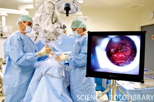
Our network hospitals have the most advanced spine surgery facilities. The spine surgeons here use minimally invasive and computer guided techniques in spine surgery. In a Minimally Invasive spine surgery the surgeon makes a few small incisions unlike the open surgery where a single large incision is made. Minimally Invasive spine surgery has several important benefits for the patients. If you have been advised spine surgery your first choice should be the latest minimally invasive technique developed by our world best hospitals in India. Most advanced Technology like Intra Operative MRI, Brain Suite and Computer Assisted Navigation System are deployed by highly trained surgeons for accurate and safe Spine Surgeries through a small incision.

Why should you choose to get Indian hospitals offer the best spinal surgery treatment in India at affordable prices. MyMedOpinion associated best spine surgery hospitals in India have the latest technology and infrastructure to offer the most advanced spine surgery at low cost.

Salient features of these hospitals are:
  • Comprehensive management of spine disorder from birth defects to degeneration of tumor and trauma.

  • Dedicated team of International trained and vastly experienced Spine Surgeons, Rheumatologists, Neurologist, Physicians and Physiotherapist.

  • Expert evaluation of spinal problems by dedicated team of experienced spine Surgeons, Rheumatologists, Neurologist, Physicians and Physiotherapist.

  • Latest Generation Diagnostic and Imaging facilities including dynamic digital X rays, Spiral CT scanning , MRI and Electrophysiology unit all under one roof.

  • Physiotherapy and Rehabilitation by experts after the surgery help you regain functional abilities quickly helping in vastly improved overall results.

Monday, 29 June 2015

Percutaneous Endoscopic Lumbar Discectomy (PELD) - Minimal Invasive Spine Surgery in india


Percutaneous Endoscopic Lumbar Discectomy (PELD)

PELD or Percutaneous Endoscopic Lumbar Discectomy is an advanced minimally invasive spine surgery done for the decompression of the lumbar disc space and removal of nucleus pulposus via a posterolateral approach. PELD is also a good treatment option in management of spinal tumors and structural deformities also in patients whose pain threshold is low or who are not responding well to conservative treatments like pain killers, anti inflammatory drugs and rehabilitation.

Percutaneous Endoscopic Lumbar Discectomy (PELD) is not the Conventional excision but a treatment which in between the conservative and open surgery. Therefore, it does not disrupt the normal soft tissue or the vertebral structures. After inserting a thin wire into the skin, the surgeons treat the herniated disc with laser and radio-frequency thermal effect under endoscopic guidance.


What is Percutaneous Endoscopic Lumbar Discectomy (PELD)?
Percutaneous Endoscopic Lumbar Discectomy (PELD) is a unique approach for the treatment of non-sequestrated disc herniation (disc prolapse). An advanced minimal invasive technique, PELD is performed for the decompression of the lumbar disc space and removal of nucleus pulposus via a posterolateral approach. In addition to decompression surgery, the technique has been found effective in management of spinal tumors and structural deformities.

What does the procedure involve? 

Percutaneous Endoscopic Lumbar Discectomy is a minimal invasive spine surgery. It is an endoscopic surgical procedure which is conducted under local anaesthesia with mild sedation. The patient is made to lie on his/her front and the exact entry point is mapped on patient’s body using image intensifier X-ray system. A small incision of about 8-10 cm is made from the midline on the posterior part of the body.
 
There are two different ways in which the inter-vertebral disc material can be removed:
  • Manual procedure also known as Automated Percutaneous Discectomy involves insertion of a long spinal needle from the side of the back, directly into the disc, by-passing other bone and ligaments. Through this needle, guide wire is passed and after making a 5 mm incision, a dilator and working cannula are passed, through which an endoscope is inserted. The prolapsed part of disc is removed with the guidance of the camera and monitor attached to the endoscope.
  • The laser surgery, referred to as Percutaneous Laser Discectomy (PLD), uses an external imager called the fluoroscope to direct the probe. The probe delivers laser energy which vaporizes a part of the inter-vertebral disc material and decompresses the nerve root. The most frequently used laser for treatment is Holmium Yttrium-Aluminum-Garnet (YAG) laser.


In either procedure, the wound is closed with single stitch. The patient gets immediate pain relief.

Duration: 60 minutes
Risks
  • Improper placement may result in nerve damage
  • Inadequate decompression may result in the need for a second operation, using a different technique
Benefits
  • Surgery is done under local anaesthesia
  • No muscle, ligament or normal tissue damage and minimal blood loss
  • No prolonged bed rest required after surgery
  • Early return to work
  • Even prolapsed, migrated, extra-foraminal, recurrent discs can be removed
  • Very good technique for old and medically compromised patients

Thursday, 4 June 2015

What about new and less invasive procedures for Back Surgery

There are three basic types of back surgery


1. A laminectomy – removing the back part of the bone (called the lamina) over the spinal column. A laminectomy is performed to relieve nerve root compression (pinched nerve) on one or more nerve roots in the spinal column. The compressed nerve root often causes back and leg pain. A segment of the entire lamina can be removed to relieve pressure on a nerve..
2. A discectomy – removing a portion of a disc to relieve pressure on a nerve.
3. A spinal fusion – this involves the permanent fusion of two or more vertebrae for more stability, to correct a deformity or to relieve pain. The surgeon will harvest small pieces of bone from your hip or pelvic bone and place them between the vertebrae. In many cases they will use wires, rods, screws, metal cages or plates to provide immediate stability.
Laminectomy
How is a laminectomy performed?

Step 1: A laminectomy is performed with the patient lying on his stomach or side and under general anaesthesia. The surgeon (an orthopaedic or neurosurgeon) reaches the spinal column through a small incision in the back.
Step 2: He will use a retractor to spread the muscles of the back apart in order to expose the bony lamina.
Step 3: He cuts away part of the lamina to uncover the ligamentum flavum – a ligament supporting the spinal column.
Step 4: In the next step he will cut an opening in the ligamentum flavum to reach the delicate spinal canal containing the compressed nerve.
Step 5: The compressed nerve can now be seen as well as the bundle of nerve fibres (known as the cauda equina) to which it is attached. It is now possible to identify the cause of compression: a bulging, ruptured or herniated disc, or perhaps a bone spur.
Step 6: The source of the pressure can now be removed. This may involve removing the bulging portion of the disc or the bony spurs and scar tissue. The herniated disc is removed after the compressed nerve has been gently retracted to one side. The surgeon will remove as much of the disc as is necessary to take pressure off the nerve. Sometimes a fragment of disc has moved and presses on the nerve root as it leaves the spinal canal. This will often cause more severe symptoms.
Step 7: With the cause of compression removed, the nerve can now begin to heal. The space created by removal of the disc will gradually fill with connective tissue. The incision is closed in several layers, from the inside outwards.
Step 8: The skin layer will be closed with steri-strips, sutures or skin clips. A dressing will be placed over the incision to protect the wound.
This operation is normally performed within one or two hours, depending on the number of levels that are decompressed.
After surgery
A plastic drain will run from inside the wound to remove any accumulating blood. In most cases, the drain can be removed on the second day after surgery.
An intravenous line to administer medication may remain connected through a vein in your hand or arm for two to three days.
Pain can and should be well controlled. Usually the acute pain subsides after a day or two. Pain may be most severe in the lower back. Leg pain may be caused by swelling of the previously compressed nerve and the trauma of the surgery. Muscle spasms across the back and down the legs are not uncommon and this can be relieved by muscle relaxants.
A physiotherapist will help you to begin standing and walking again, and show you how to get in and out of bed and how to sit, stand, and sleep.
 What about new and less invasive procedures?
The era of less invasive surgery has dawned – also in back surgery. Smaller keyhole incisions are replacing large surgical cuts. These less invasive techniques include:

  • Endoscopic discectomy – this technique employs a disposable scope that the surgeon inserts through a small incision. The protruded disc that is compressing the nerve is then removed using specially designed instruments.
  • Vertebroplasty – the injection of bone cement into a fractured vertebrae. The hardened cement will seal and stabilise the fracture and relieve pain. People suffering from severe pain because of a compression fracture will be considered for this procedure, especially if they have difficulty standing and walking.
  • Kyphoplasty – the insertion of a “balloon” to expand a compressed vertebra and the injection of bone cement


  • MyMedOpinion  affiliated   Best hospitals in India provide an medical opinion from experienced surgeons and the treatment cost includes companion stay  , surgeon fee, medicines and consumables, nursing care, patient's food and airport pick up & drop etc. etc. We offer free, no obligation assistance to international patients to find world class medical treatment in India. We offer support and services to facilitate the care you require. We can help you find the best hospital in India

    Send us a Medical Report to Get FREE Medical Opinion from India's Top Doctors


    MyMedOpinion is the hub of Surgeons and specialists for major diseases. We give you a chance to speak to our doctors and discuss your health issues directly.

    Email Us: Free@mymedopinion.com  Call Us: +91-7503537224